Category: PT Corner with Kristin Williams

  • The Almighty ACL

    The Almighty ACL

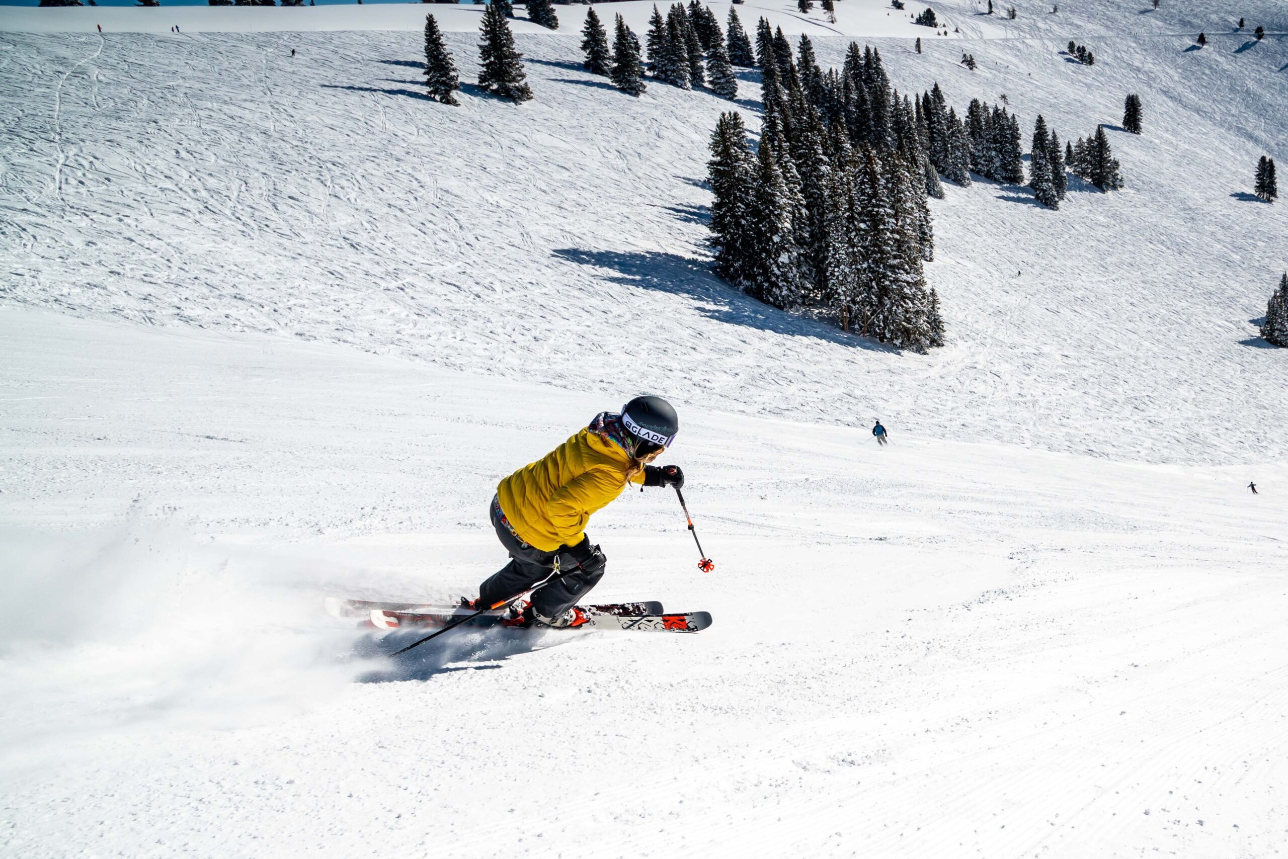
    With ski season just around the corner, it’s time to talk about the almighty ACL. I used to love to ski with abandon until I became a physical therapist and then skiing was never the same again! ACL sprains are common knee injuries and skiers are one of the most at-risk athletes. Others include soccer, basketball, and football players. That being said, I can no longer go downhill skiing without an arguably irrational fear of tearing or spraining my ACL. Sigh. 


    The ACL, which stands for anterior cruciate ligament, is one of two cruciate ligaments that aid in stabilizing the knee joint. The ACL and PCL (posterior cruciate ligament) form an “X” or cross within the knee. The ACL comes from the posteromedial of the medial aspect of the lateral femoral condyle and inserts on the anteromedial tibia, blending with the medial meniscus. As it passes from the femur to the tibia, it spirals outward (laterally) on itself. 

    anatomy-of-the-knee


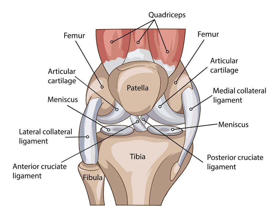
    The primary function of the ACL is to restrain the tibia (shin bone) from moving forward on the femur (thigh bone). It also restrains rotation and medial/lateral (varus or valgus) displacement of the tibia when the knee is straight. Finally, the mechanoreceptors present in all ligaments provide crucial proprioceptive information to the brain as to where the knee is in space at any given time.


    Roughly 70% of all ACL injuries are caused simply by doing the wrong movement. Furthermore, approximately 75% of all ACL ruptures are sustained with minimal or no contact. A common mechanism of injury involves a cut-and-plant movement, especially if there is rapid deceleration involved, which is why skiers are at-risk for injury. When turning, if there is a sudden change in direction of the knee but the foot stays firmly planted in the ski which is still pointing downhill, you’re replicating the cut-and-plant movement and can tear the ACL. Other mechanisms of injury include rapid stopping, landing awkwardly, pivoting, twisting, and direct blows to the leg.


    Studies have shown that females are at increased risk of ACL injury and have a 2.4-9.7 times higher incidence rate as compared to males. Some studies suggest that females have weaker hamstrings, so they mostly utilize the quadriceps while decelerating. This places increased stress on the ACL, as the quads are less effective at preventing forward movement of the tibia as compared to the hamstrings. A second factor that may increase the risk in females is a wider pelvis requiring the femur to have a greater angle towards the knee. One study using video analysis demonstrated that female athletes are more likely to bring the knees in when changing directions suddenly (creating a valgus stress), which increases the strain on the ACL. Finally, hormonal variations may increase the laxity of ligaments overall in females.


    When someone sustains an ACL tear, there may be an audible pop and the knee will often give out underneath them, which is often followed by an immediate onset of pain and significant swelling. Range of motion may be restricted, especially full extension. There may be widespread mild tenderness to touch as well. If not repaired, episodes of giving way are common, especially with pivoting and twisting motions. People are able to function without an ACL, but this often leads to an earlier onset of arthritic changes to the joint due to the decreased stability and increased wear and tear. 


    They say an ounce of prevention is worth a pound of cure, which has never been more true than with the ACL. An ideal ACL prevention program will incorporate plyometrics, neuromuscular training, muscle strengthening, and education on body mechanics and landing technique. Plyometrics should include high intensity agility drills that develop footwork and power, including cutting, jumping, and lateral movements. The athlete should be taught to begin and end movements with proper positioning involving hips and knees being sufficiently flexed, jumping and landing with knees over the foot avoiding a valgus stress, and landing softly. Strength training should be focused on the hamstrings and glutes. Any asymmetry in strength and movement patterns should also be addressed. Hmmm…does any of this sound familiar?? LYT Yoga® fits the bill and is the perfect preseason prevention training! However, if you find yourself struggling from an ACL sprain, click the link below to our YouTube channel for some simple exercises to help rehabilitate your knee following an injury. Until then, I’ll see you on the mat!


    Xoxo,

    Kristin

  • Warming Up to the Cold

    Warming Up to the Cold

    It’s getting to be that time of year where everyone is looking to get warm as the temperatures drop. Ever since moving to the tundra of Wisconsin and dealing with the winds of the lake effect, my attention to staying warm despite the cold has become hyper-focused! As the temperature outside decreases, body heat is lost more quickly and it becomes more of a challenge to maintain a normal body temperature. Luckily we have some good defensive mechanisms in place to help us stay warm when baby, it’s cold outside!


    The first line of defense occurs right when we encounter colder temperatures and our body automatically redistributes blood to the torso in order to keep the vital organs warm and in working order. The blood vessels of the skin constrict minimizing the amount of blood sent to this area, which is the closest to the cold. This helps to insulate the body by keeping the heat from escaping and shunts the warm blood to the core. Another strategy the body uses to warm up is shivering. You know when your teeth begin to chatter and your body starts to shake uncontrollably? It’s all for a good reason. These rapid and rhythmic muscle contractions create heat in order to keep the body warm. The body may also recruit more muscles to shiver as the temperature drops, so it can actually become quite intense and even uncomfortable. This is an effort by the body to increase body temperature by breaking down nutrients and maintaining the feeling of warmth. In fact, shivering can increase heat production close to five times the resting metabolic rate! 


    So why is it that some people get cold more easily than others? Body type can explain some of the varying reactions. Taller people tend to get cold faster because a larger surface area means more heat loss. In addition, fat’s reputation as an insulating material is well deserved, so thinner people also tend to get cold quicker. It’s like a double whammy for those who are both tall and thin! For staying warm during the winter, subcutaneous fat that is layered under the skin is what keeps the heat in, not the visceral fat that collects in the abdomen. This is why inuit and other polar/cold climate peoples have evolved to be relatively short and stout. 


    So what’s a great way to warm up to the cold? Simply being physically active is a great way to generate heat. During exercise, heat is created as your muscles use and create energy. Typically, a muscle stores a small amount of energy within, and once this has been used, it must create additional energy to continue working. In fact, when you exercise, 80% of the total energy is converted to heat while only 20% is used for muscle contraction. Exercise is not a total gain though, because once you experience an increase in body temperature, your body will start sweating in an effort to cool down. In cold temperatures, this can become problematic as the evaporation of sweat can lead to greater rates of heat loss. This is why proper clothing that allows for wicking of moisture away from the skin while also insulating the body to keep warm is imperative for outdoor cold weather exercise.


    Therefore, now that the temperatures are starting to fall, we have even more reason to get on our mats and keep moving! Wear some layers that you can shed as your body temperature rises and let LYT Yoga® loosen up whatever Old Man Winter has tightened! On that note, I’ll see you on the mat!


    Xoxo,

    Kristin

  • Carpal Tunnel

    Carpal Tunnel

    With more people working from home and online than ever, I wouldn’t be surprised if we see an uptick in the number of carpal tunnel syndrome (CTS) cases worldwide. CTS is the compression of the median nerve as it passes through a fibrous tunnel at the wrist into the hand, resulting in pain, numbness, tingling, and weakness along the distribution of that nerve. It is the most common nerve entrapment syndrome, accounting for 90% of all entrapment neuropathies. Given that it is made worse by prolonged extension of the wrist, one can understand why activities such as using a mouse and typing would aggravate symptoms of CTS.


    Developing CTS is often multifactorial, involving occupational, social, and environmental risk factors. The most common risk factors include genetic predisposition, history of repetitive wrist movements, obesity, autoimmune disorders, and pregnancy. CTS is 10 times more common in females than males and usually occurs between the ages of 40-60 years. It is most often caused by a combination of compression and traction at the wrist. With compression, there is a cycle of increased pressure > obstruction of blood flow > increased swelling > compromise to the microcirculation of the median nerve > compromise of nerve function > lesions in the nerve itself > inflammation of the surrounding connective tissues and tendons passing through the tunnel > further compression of the nerve. Repetitive traction and wrist motion can only exacerbate symptoms, further injuring the nerve.


    Sensory nerve fibers are often affected before motor fibers, resulting in early symptoms of pain, numbness, and tingling. These symptoms will present along the distribution of the median nerve, which includes the thumb, index finger, middle finger, and half of the ring finger (closest to the thumb). Pain can also radiate up into the arm. As the disease progresses, motor nerve fibers are affected, causing hand weakness, decreased fine motor skills, and atrophy of the muscles of the thumb. Autonomic nerve fibers can also be affected, causing temperature changes in the hand. 


    In the early stages, symptoms are most often present at night when lying down and decrease during the day. Symptoms at this stage can often be relieved by shaking the wrist and hand. As the nerve entrapment progresses, symptoms will also be present during the day, especially with repetitive wrist and hand activities. In more advanced cases, the symptoms can be constant. As CTS is progressive, permanent median nerve damage can result. However, almost 90% of mild to moderate cases respond to conservative management.


    Three simple tests to check for CTS include:

    1. Carpal Compression Test – most reliable – done by applying firm pressure directly over the carpal tunnel for 30 seconds. The test is positive if pain, numbness, tingling, or other symptoms are reproduced.
    2. Phalen Test – aka “Reverse Prayer” – Fully flex the wrists, placing the backs of the hands together for one minute. A positive test is when the symptoms are reproduced.
    3. Reverse Phalen Test – aka “Prayer Test” – Fully extend the wrists, placing the palms of the hands together for one minute. A positive test is when the symptoms are reproduced.


    As I stated above, conservative treatment of CTS is highly successful in a lot of cases. Initially, just modifying movements or positions that provoke the symptoms is key to breaking the compression cycle. Increasing aerobic activity, mobilizing the upper thoracic spine, stretching tight muscles of the cervical spine and thoracic outlet, strengthening the shoulder girdle, and gliding the median nerve can also help decrease aggravating factors of CTS. I’ve created a short video that can be found on our LYT Yoga® YouTube channel with simple exercises you can do to help relieve the symptoms of CTS, so check out the link below! Until then, I’ll see you on the mat!


    Xoxo,

    Kristin

  • Posture Matters

    Posture Matters

    There are a lot of healthcare professionals on social media discussing whether or not posture really matters. It’s kind of the hot button topic in the physical therapy and pain science world. These days, it’s en vogue and edgy to say that posture doesn’t matter. I’ve been in this business long enough now to watch fads come and go. I love to listen to both sides of every story and to be honest, most of the time the underlying message on each side is essentially the same. People are just too busy spewing out extreme messages to get more likes on their Instagram page rather than really listening to what others have to say. Since this is my platform to educate people about the body as best I know how, I want to tell our readers why I think posture matters…but it may not be in the way you’d expect.


    When most people think of good posture, they think head up, shoulders back, don’t slouch, and suck in your stomach, perhaps like an Army PFC lined up for uniform inspection. Is that what we want? Is that how we should present at all times in order to avoid back or neck pain? No. But does that mean it doesn’t matter? Does that mean slouching with a forward head and neck, zero tension in the core, and a tilted pelvis is ok? Again, no. Posture matters in the sense that being aware of where your body is in space at all times matters. There is little to no solid evidence that having bad posture causes pain. In fact, there is a lot of evidence to the contrary…that poor posture does not cause pain. But that doesn’t mean it doesn’t contribute or play a role. One study looked at thousands of people who experienced an episode of low back pain and found that being distracted while doing a specific task makes people 25x more likely to experience acute low back pain. It also found that being in an awkward posture also increased the likelihood, just not as much. The combination of the two, an awkward posture and being distracted, was the key. 


    There is also evidence that posture affects emotions. After all, you rarely see a superhero portrayed in a rounded, traditionally poor position of posture. People in positions of power often adopt a similar position for that same reason. Power begets power. A depressed posture has been shown to cause depression. Depressed people who adopt happy postures have been shown to feel better. There is also evidence that emotions affect pain sensitivity. Anxiety increases perceived pain responses. So it stands to reason that posture, emotions, and pain are related in some fashion and clearly influence one another. 


    It’s better to think of posture and movement patterns in terms of what puts the most amount of postural stress on the body. Younger people with more adaptable soft tissues and mobile joints may be able to maintain an awkward or poor posture for longer periods of time than an older person with less adaptable tissues because the postural stress on the body is less overall. Duration of stress matters as well. Bending over to examine something under your sink for five minutes is much less likely to cause injury as opposed to doing that for many hours a day as a plumber, for example. The duration of the stress is longer, but the posture is the same. Many postural stresses can be avoided…if you notice it’s a stressor…which unfortunately, many people don’t.


    The best posture is a dynamic one. We weren’t created to be sedentary. Our bodies are meant to move in a variety of positions and to do so frequently. A sedentary lifestyle contributes significantly to the degeneration of postural reflexes, as discovered by NASA while studying the physiological effects of inactivity. We have the best length-tension relationship in our muscles in neutral, so it’s a great place to start. Once you’re able to identify neutral, your brain and your body become more aware of when you stray and it’s easier to respond as appropriate, for the position you find yourself in. You don’t and shouldn’t maintain erect Triple S posture throughout every movement. Find freedom and variety in your movements, with the ability to decrease the postural stress as needed for your body at that specific time. It’s a learning process that takes time and practice. And it matters. 


    Check out the link below to our Posture Series on LYT Daily. It’s also available for purchase if you aren’t a subscriber. In this series, Lara and I educate you, your brain, and your body on all things posture and movement, to decrease those postural stressors in your life! Until then, I’ll see you on the mat!


    https://lytyoga.uscreen.io/programs/collection-9eqop9lxcpu


    Xoxo,

    Kristin

  • Collagen

    Collagen

    I never used to think much about collagen until I got into my 40’s. Now I can’t help but hear about it almost every day in ads for skin and joint health. So what’s the big deal that makes collagen such a hot topic in healthcare and beauty? In LYT Yoga®, we’re constantly talking about “soft tissues” and “connective tissues”. Similar to the framework of a house, connective tissue provides structure, support, and protection throughout the body. Connective tissue is comprised of ground substance, fibers, and cells. The ground substance and fibers make up something called the extracellular matrix, which is the structural support of connective tissue. The three types of fibers include elastin, reticulin, and of course, collagen.


    Collagen fibers are large and strong, providing high tensile strength to the extracellular matrix and can be found in both dense and loose connective tissue. There are actually 28 different types of collagen in the body, with four being the most common:

    • Type I – makes up over 90% of the collagen in the human body – found in all connective tissue, but most notably scar tissue, tendons, ligaments, bone, skin, eyes and teeth. It’s both flexible and strong, providing resistance to tension, force, and stretch.
    • Type II – found in the cartilage of joints and intervertebral discs, providing resistance to pressure.
    • Type III – often found in organs such as skin and blood vessels, providing a flexible meshwork for cells – it’s also abundant during the early stages of wound healing.
    • Type IV – an essential component to the membranes of the kidneys, inner ear, and lens of the eye, providing support and sites of attachment.


    As we age, the production of collagen begins to slow, which causes the cell structures to weaken. Skin becomes thinner, drier, and less elastic, causing wrinkles to form and skin to sag. Womp womp. Bones become weaker and more fragile. Ligaments lose their elasticity. Joints become stiffer as the cartilage wears down. Finally, muscle function decreases. Woof.


    But don’t lose hope! Exercise has been shown to prevent cartilage degeneration in the body and help keep the skin healthy and vibrant. Researchers have shown that during exercise, the compression of the joint cartilage (made up of collagen) stimulates the tissue cells to block the action of inflammatory molecules associated with conditions like arthritis. There is certainly a threshold of doing enough, but not too much however. Exercise also increases blood flow, which helps to nourish skin cells. In addition to providing oxygen, blood carries away waste products, including free radicals, from cells. So you can think of it as helping to flush the system.


    So the next time you hop on your mat and your favorite LYT Yoga instructor is challenging you to work a little harder or sweat a little more, think of the good you’re doing for your connective tissues and collagen! 🙂 If you want more information on healthy skin care, check out our LYT Yoga Blog by Jana Broeckx, European LYT Yoga teacher and entrepreneur! See the link below! You know we’re always pulling for you! Until then, I’ll see you on the mat! 


    Jana’s skincare story


    Xoxo,

    Kristin

  • Pes Planus (Flat Feet)

    Pes Planus (Flat Feet)

    If I had a dollar for every person who told me during their past medical history interview that they have “flat feet”, I’d have a small nest egg in the bank. Most of the time when I do my examination however, I see nothing of the sort. Especially nowadays when shoe manufacturers have capitalized on “over-pronation”, many people are under the impression that they have flat feet when they really don’t. In fact, truly flat feet are rare.

    flat foot

    Pes planus, or flat feet, can be either congenital or acquired and is defined by the loss of the medial longitudinal arch of the foot. The arch of the foot is a tough, yet elastic combination of ligaments, tendons, and fascia connecting the forefoot to the hindfoot. It acts as an adaptable and flexible base of support for the entire body, dissipating the forces of weight-bearing and storing mechanical energy during the gait cycle. Dysfunction of any portion of the medial longitudinal arch can result in an acquired pes planus. Risk factors for developing flat feet include excessive tension on the gastroc-soleus complex (the calf muscles), obesity, ligamentous laxity, or posterior tibialis tendon dysfunction. The function of the posterior tibialis tendon is to support the arch as well as invert and point the foot. A failure or dysfunction of the tendon can contribute to a fallen arch in weight bearing. Posterior tibialis dysfunction is most common in females over the age of 40 who have other chronic health issues such as diabetes and obesity. It can also occur in people who have congenital pes planus who participate in repetitive high impact sports.


    It’s not uncommon for toddlers and young children to have flat feet due to ligamentous laxity and a lack of neuromuscular control. However, most children develop normal arches by age 5 or 6. It is a small percentage of children who fail to develop a normal arch by adulthood, making the percentage of people with truly collapsed arches, or rigid pes planus, relatively small. It is estimated that between <1-28% of the population has some degree of pes planus, but a majority of these cases are flexible pes planus. With flexible pes planus, the arch is present in non-weight bearing, but absent or decreased in weight bearing. Rigid pes planus is rare. There is a strong genetic component of pes planus, so it typically runs in families. People with congenital ligamentous laxity secondary to Down syndrome, Marfan, or Ehlers Danos can present with flat feet. The ligamentous laxity associated with pregnancy can also cause flat feet, but typically corrects itself postpartum. 


    The main symptom of pes planus is foot pain due to strained muscles and connective tissues. People may have pain along the posterior tibialis tendon or with a single leg heel raise. If the collapse of the arch is severe, the ankle may turn inwards and the bulk of the body weight is placed through the medial border of the foot. Such a distortion of weight bearing often results in abnormal biomechanics of the lower extremities and can cause calf, knee, hip, or low back pain.


    Treatment of pes planus includes increasing the flexibility of the feet and lower legs, strengthening the small muscles of the foot, lower legs, hips, and core, training proprioception, and patient education. LYT Yoga® certainly ticks all of these boxes! In fact, if you want to learn about all things feet, Lara has an upcoming workshop entitled “Foundations of the Foot” on October 30 that you won’t want to miss! Check out the link below! Until then, I’ll see you on the mat!


    https://lytyoga.uscreen.io


    Xoxo,

    Kristin

  • Shoulder Labral Tear

    Shoulder Labral Tear

    Many of you who practice with me regularly know that I have a cranky right shoulder. Several years ago, I was working on a fellow physical therapist who is around 6’2” and roughly 250 lbs. His back was bothering him so I wanted to help him out. It had been a long morning of treating patients. I began to manipulate his sacroiliac joint, a maneuver that requires me to thread my arm through his. In this maneuver, my right shoulder was flexed and internally rotated.


    I’ve done this technique thousands of times without any issue, but this time when I leaned away and used my body weight to roll him, I felt and heard a tearing in my shoulder. He heard it too.  He looked at me, I looked at him, and we simultaneously muttered, “Uh-oh.” I never had any diagnostic tests done and rehabbed it myself, but I suspect I tore my labrum.


    The labrum of the shoulder is a fibrocartilaginous rubbery rim attached around the margin of the socket. It cushions the joint called the glenoid fossa, which is part of the scapula (shoulder blade). The socket is quite shallow and small, covering at most only a third of the ball (the head of the humerus). 


    One function of the labrum is to deepen the socket. It increases the contact area between the ball and socket by 2 mm at the front and back and 4.5 mm at the top and bottom. If you took a cross-section of the labrum, it would look like a triangle, where the wide base attaches to the edge of the socket, leaving the edge of the labrum thin and sharp at the point. This shape allows it to almost act like a washer, sealing the ball and socket together. 


    This is called a “viscoelastic piston effect” and maintains a negative pressure within the joint. It’s especially effective against traction or pulling stresses and, to a lesser extent, against shearing stresses. Finally, the labrum provides an insertion point for stabilizing structures, including the joint capsule ligaments and the tendon of the long head of the bicep muscle.


    The labrum is often compared to the face of a clock, with 12 o’clock being at the top (superior), 3 o’clock at the front (anterior), 6 o’clock at the bottom (inferior), and 9 o’clock at the back (posterior). Most instabilities in the shoulder are associated with injuries to or changes within the glenoid labrum, particularly where the long head of the biceps tendon inserts at the superior (12 o’clock) portion. They are commonly called SLAP tears, as they involve a Superior Lesion from Anterior to Posterior, usually between 10 and 2 o’clock.


    Different types of SLAP tears typically involve different mechanisms of injury.  A common mechanism is falling on an outstretched arm or pulling suddenly when lifting a heavy object, as I did with my fellow PT. Other mechanisms include repetitive shoulder abduction and external rotation, like the movements that many throwers, overhead athletes, or manual laborers perform over and over.  A direct blow to the shoulder, as with tackling in football can also be a cause. SLAP tears can occur in a degenerative manner for the aging population as well.


    Treatment for SLAP tears includes both nonoperative and operative measures. Both have proven successful among certain populations. Previous studies indicate that nonoperative management is successful for anywhere from 22 to 85% of patients. Operative repair in adults is reported to be successful for between 80 and 97% of patients. It often helps alleviate the pain and return of range of motion.  


    Among overhead athletes, many patients are unable to return to their prior level of performance.  Results vary widely in this population, with between 7 and 84% demonstrating a return to their prior level of performance. As patients age beyond 40 years old, surgeons typically opt to perform a biceps tenodesis. In this surgical procedure, the tendon of the long head of the bicep is detached from the labrum and anchored to the proximal humerus. Repair success rates are lower among older populations, so this is the most effective treatment of choice.


    I continue to address my shoulder issue with home exercises and activity modifications and so far, so good! As I say all the time in Stretch class, the shoulder joint is our most mobile, but also our most unstable. For all the parts to work in harmony, it requires a high quality of movement.  Paying attention to how we move on our mats and keeping the ball centered in the socket is important for many reasons, but especially to avoid placing repetitive undue strain on the labrum.


    Continue using your LYT Yoga® practice to build strength and good brain mapping in the shoulder joint complex. Until then, I’ll see you on the mat!


    Xoxo,


    Kristin

  • Vestibular System

    Vestibular System

    Proust quote

    ~ Marcel Proust


    Lara’s quote this week references looking at the world through new eyes and it made me think of how disorienting the world is if something messes with our vision! For example…vertigo. If you’ve never had it, lucky you! If you have, you are fully aware how your vision can be affected by it.


    Vertigo is caused by a disruption to the vestibular system, which is a complex set of structures and neural pathways that helps us maintain balance and spatial orientation. It does so by detecting the position and movement of our head in space. There is a vestibular apparatus in the inner ear where this whole process begins. It sends signals to multiple places in the body, including the eyes and central nervous system. This is all reflexive, or unconscious. The activity between the vestibular system and the eyes is called the vestibulo-ocular reflex (VOR), which allows the eyes to remain fixed on an object while the head is moving. The activity between the vestibular system and the spine is called the vestibulospinal reflex (VOS), which coordinates the muscles of the spine with head movement to maintain balance and posture.


    Any disruption along the pathway can lead to various symptoms including vertigo, imbalance, nausea, vomiting, visual disturbances, hearing changes, and uncoordinated eye movements. When you have vertigo, it can feel like the world is spinning despite the fact that you’re lying completely still. As a result of this vestibular disruption, dizziness, nausea, and vomiting can occur. The most common type of vertigo is benign paroxysmal positional vertigo (BPPV), which usually only lasts for seconds to minutes. It is believed to be due to the displacement of tiny crystals in the ear canals called otoconia which causes an inappropriate sensation of movement. I suffer from occasional bouts of BPPV, which luckily only last a couple of seconds, but can be very disorienting. In my case, when I turn my head, my vision stutters hard back and forth for a second or two and I feel a sudden onset of dizziness. It goes away as quickly as it comes on, but can leave me feeling a little nauseated. Most people report having a spinning of their visual field, which often causes a loss of balance coupled with nausea and/or vomiting. A hallmark of BPPV is nystagmus, which is uncoordinated eye movement in response to moving the head. So when a person’s head is turned in one direction, the eyes will beat quickly back and forth, indicating a failure of the VOR to work.


    Meniere disease is another type of vertigo which can last for hours with the same symptoms as BPPV, but also includes hearing loss and tinnitus, or a ringing in the ears. With Meniere, there is an expansion of fluid in the ear, impacting the vestibular apparatus. My dad suffers from Meniere disease, so clearly vertigo runs in the family! In his case, he will typically have it right upon waking in the morning and he immediately feels nauseated. If he tries to get out of bed, he will have violent episodes of visual spinning and vomiting. It can last for the entire day. By the next day, although tired, he usually feels fine.


    Another type of vertigo is viral labyrinthitis, which is caused by inflammation of the vestibular nerve as a result of a viral infection. Symptoms can last from days to weeks and include hearing loss in the ear that is affected and a loss of balance. These three forms of vertigo are called peripheral because they include the inner ear as the main source of vestibular disruption. There are also forms of vertigo caused by central nervous system lesions in the brainstem, pons, or cerebellum, which are much more serious in nature. Central vertigo can be the result of a stroke, multiple sclerosis, toxicity due to medication, tumors, etc.


    It’s not until you lose your vision that you really appreciate its role not only in daily function but in balance and proprioception. In LYT Yoga, I like to create classes that challenge the brain and body by purposely affecting the vestibular system through movements of the head or closing the eyes. It not only heightens our appreciation of this intricate and vital ability, but helps to train each individual portion, making us stronger in the long run. Click the link below to check out my class “Toe The Line” and see how your vestibular system is working! Until then, I’ll see you on the mat!


    https://lytyoga.uscreen.io/programs/toe-the-linemp4-6cb992


    Xoxo,

    Kristin